Pinhole Imaging and Lens Imaging
Imaging refers to the technique of forming images of biological samples, which can be broadly divided into tissue imaging and cellular molecular microscopy techniques based on the scale of the samples. These techniques generally require the development of optical technologies in conjunction with the characteristics of biological samples, with some employing wave properties other than light, such as nuclear magnetic resonance and ultrasound.
Pinhole Imaging
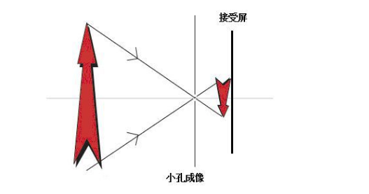
When a board with a small hole is placed between a screen and an object, an inverted image of the object is formed on the screen. This phenomenon is called pinhole imaging. Moving the board back and forth will change the size of the image. This phenomenon reflects the property of light propagating in straight lines.
Pinhole imaging dates back to approximately 2,500 years ago in ancient China when the scholar Mozi (Mo Di) and his students conducted the world's first experiment on pinhole imaging, explaining the reasons for the inverted image formation and pointing out the property of light propagating in straight lines. This was the first scientific explanation of the propagation of light in straight lines.
Pinhole Imaging Experiment
-
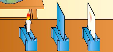
Arrange the candle, pinhole screen, and ground glass screen as shown in the diagram. Light the candle and adjust the height of the candle and the screen so that the flame of the candle, the pinhole, and the center of the ground glass screen are roughly aligned on a straight line. The distance between the candle and the pinhole screen should not be too large. After adjustment, an inverted real image of the candle flame can be seen on the ground glass screen.
-
Moving the candle or the position of the ground glass screen, it can be observed that the closer the candle is to the pinhole or the farther the ground glass screen is from the pinhole, the larger the image obtained.
Principles Obtained
- As long as the pinhole is small enough, regardless of its shape, it has little effect on the clarity and shape of the resulting image.
- The closer the object is to the pinhole, the smaller and brighter the resulting image; conversely, the farther the object is, the larger and dimmer the image.
- The closer the pinhole is to the candle, the larger and dimmer the resulting image; conversely, the farther the pinhole is, the smaller and brighter the image.
- In the pinhole imaging experiment, the resulting image is an inverted real image, and the size and clarity of the image are related to the conclusions above.
Lens Imaging
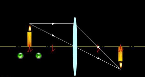
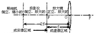
Lenses are divided into convex and concave lenses. The imaging law of convex lenses is as follows: when the object is placed outside the focal point, an inverted real image is formed on the other side of the convex lens, which can be either reduced, equal-sized, or magnified. The smaller the object distance, the larger the image distance, and the larger the real image. When the object is placed inside the focal point, a virtual upright magnified image is formed on the same side of the convex lens. The larger the object distance, the larger the image distance, and the larger the virtual image. Concave lenses diverge light rays and have a much more complex imaging law.
In optics, an image formed by actual light rays converging is called a real image, which can be captured by a screen; conversely, an image formed by diverging light rays is called a virtual image, which can only be perceived by the eyes. When experienced physics teachers explain the difference between real and virtual images, they often mention a method of distinction: "Real images are always inverted, while virtual images are erect." The terms "erect" and "inverted" are of course relative to the original object.
The three types of virtual images formed by plane mirrors, convex mirrors, and concave lenses are all upright; while the real images formed by concave mirrors and convex lenses, as well as the real images formed in pinhole imaging, are invariably inverted. Of course, concave mirrors and convex lenses can also produce virtual images, and the two types of virtual images they produce are also in an upright state.
So, are the images formed by human eyes real or virtual? We know that the structure of the human eye is equivalent to that of a convex lens, so the images formed by external objects on the retina must be real images. According to the above empirical rule, the image on the retina seems to be inverted. However, any object we usually see appears to be upright. This conflicting problem with the "empirical rule" actually involves the adjustment function of the cerebral cortex and the influence of life experience.
When the distance between the object and the convex lens is greater than one focal length but less than twice the focal length, an inverted image is formed, which is a convergence point of actual light rays passing through the convex lens and can be captured by a screen, making it a real image. When the object approaches the lens from a distance, the image gradually becomes larger, and the distance from the image to the lens also gradually increases; when the distance between the object and the lens is less than the focal length, a magnified virtual image is formed.
FALenses Technology specializes in providing machine vision core hardware. You can go to the official website of FALenses Technology at https://www.falenses.com/ for more information.